Bap1 Mesothelioma Immunohistochemistry : Mtap Immunohistochemistry Is An Accurate And Reproducible Surrogate For Cdkn2a Fluorescence In Situ Hybridization In Diagnosis Of Malignant Pleural Mesothelioma Modern Pathology - bap1 immunohistochemistry and p16 fluorescence in situ hybridization (fish) have recently been reported as reliable markers of malignancy in biopsies of mesothelioma.
Bap1 Mesothelioma Immunohistochemistry : Mtap Immunohistochemistry Is An Accurate And Reproducible Surrogate For Cdkn2a Fluorescence In Situ Hybridization In Diagnosis Of Malignant Pleural Mesothelioma Modern Pathology - bap1 immunohistochemistry and p16 fluorescence in situ hybridization (fish) have recently been reported as reliable markers of malignancy in biopsies of mesothelioma.. Genomic studies of malignant mesothelioma (mm) have identified frequent mutations in brca associated protein 1 (bap1), a nuclear deubiquitinase and transcriptional regulator. Formerly called atypical spitz tumors), and the following cancers, in descending order of frequency: Cautions loss of bap1 expression as determined by immunohistochemistry will not be able to differentiate between germline and somatic mutation. A combination of mtap and bap1 immunohistochemistry in pleural effusion cytology for the diagnosis of mesothelioma: She had a strong family history of mesothelioma as well as other malignancies including renal cell carcinoma.
To determine how useful these tests are with sarcomatous and desmoplastic mesotheliomas, we examined 20 such tumors. bap1 immunohistochemistry (ihc) in representative malignant pleural mesothelioma (mpm) and reactive mesothelial hyperplasia (rmh) cases. Conclusions.— in the context of a mesothelial proliferation, loss of mtap staining is 100% specific for malignant mesothelioma. Roc analysis of the two tests. Malignant mesothelioma, bap1 immunohistochemistry, and vegfa:
Pdf Malignant Mesothelioma Bap1 Immunohistochemistry And Vegfa Does Bap1 Have Potential For Early Diagnosis And Assessment Of Prognosis from i1.rgstatic.net Henderson,1,2 and sonja klebe1,2 1department of anatomical pathology, flinders university of south australia, bedford park, sa 5042, australia No cases showed loss of nf2. immunohistochemistry performed on the first pleural effusion in retrospect showed loss of bap1 at that time; Immunohistochemical and molecular testing for diagnosis of mesothelioma will be reviewed. Pulford e, huilgol k, moffat d, et al. immunohistochemistry of presumed cancer of unknown primary specimen, allowing the allocation to epithelioid mesothelioma and revision of diagnosis: Two cases were excluded from bap1 analysis: 4a) and in tumor cells of mm tissue specimens with the wild‐type bap1 (mm26, mm67, mm16, mm30, mm80, and mm46;
Differentiating malignant pleural mesothelioma from benign reactive mesothelial processes can be quite challenging.
Does bap1 have potential for early diagnosis and assessment of prognosis? immunohistochemistry performed on the first pleural effusion in retrospect showed loss of bap1 at that time; To determine how useful these tests are with sarcomatous and desmoplastic mesotheliomas, we examined 20 such tumors. A combination of mtap and bap1 immunohistochemistry in pleural effusion cytology for the diagnosis of mesothelioma. Review of atypical mesothelial proliferations versus mesothelioma from a pathologist point of view. bap1 assessment using immunohistochemistry on needle biopsy may benefit preoperative risk stratification and guide treatment planning. T, p.arg385*) in bap1 or a recurrent pathogenic germline mutation (c.301g > Peritoneal malignant mesothelioma shows weaker association with asbestos exposure and more likely involves women / young patients than pleural malignant mesothelioma. Prescreening of their diagnostic archival biopsy tissues; To determine whether these markers, singly or in combination, might also be useful in effusion cytology specimens, we examined 15 biopsies of epithelial mesotheliomas and 3 benign. immunohistochemistry (ihc) is a reliable technique to determine bap1 molecular status. We previously reported the use of. bap1 immunohistochemistry (ihc) in representative malignant pleural mesothelioma (mpm) and reactive mesothelial hyperplasia (rmh) cases.
immunohistochemistry of presumed cancer of unknown primary specimen, allowing the allocation to epithelioid mesothelioma and revision of diagnosis: The recently described bap1 hereditary cancer predisposition syndrome was suspected, but immunohistochemical labeling was not conclusive. Loss of one or the other marker was observed in 17/20. Had bap1 labelling been performed in addition, since none of the metastatic lung adenocarcinomas showed loss of nuclear labelling for bap1, loss of nuclear labelling may be an additional indicator supporting a diagnosis of mesothelioma. To determine whether these markers, singly or in combination, might also be useful in effusion cytology specimens, we examined 15 biopsies.
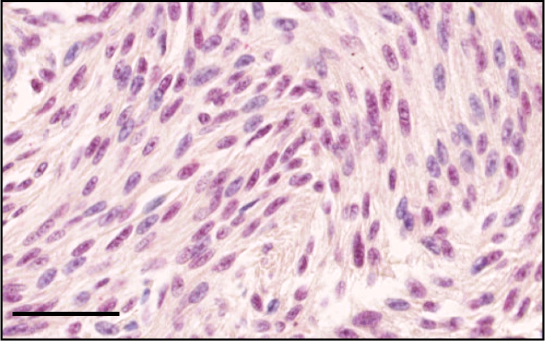
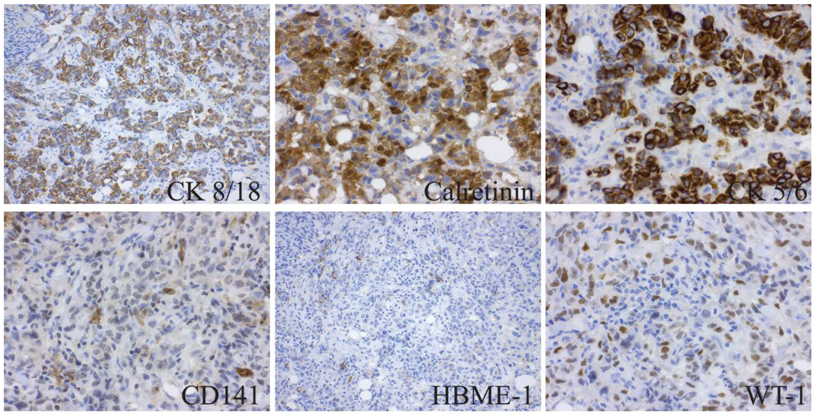
Pathology Outlines Bap1 from www.pathologyoutlines.com immunohistochemistry (ihc) is a reliable technique to determine bap1 molecular status. Histologic variants (epithelioid, biphasic and sarcomatoid) impart prognostic information with treatment implications. Interestingly, bap1 mrna transcripts in bap1 del tumors were expressed at lower levels as compared to those in bap1 intact tumors (p value = 3 × 10 −4) (fig. immunohistochemistry in patients with mesothelioma, validating loss of expression in independent international cohorts. Differentiation of malignant pleural mesothelioma (mpm) from benign mesothelial proliferation remains problematic. With respect to bap1 immunohistochemistry, 12 of 19 interpretable mesothelioma cytology cases demonstrated loss of bap1 (63%). Malignant mesothelioma, bap1 immunohistochemistry, and vegfa: Detected using immunohistochemistry (ihc)) and homozygous deletion (hd) of p16 (detected using fluorescence in situ hybridization (fish)) are useful for differentiation of mpm from reactive mesothelial hyperplasia.
4a) and in tumor cells of mm tissue specimens with the wild‐type bap1 (mm26, mm67, mm16, mm30, mm80, and mm46;
Removing ubiquitin modifiers, which alter the function of other proteins via targeted degradation, subcellular localization, or activity (1). bap1 immunohistochemistry is a useful test in many settings, including screening for germline bap1 mutations, diagnosis of malignant mesothelioma and prognostic stratification of various cancer types including uveal melanoma, cutaneous melanoma, mesothelioma and clear cell renal cell carcinoma. immunohistochemistry with anti‐bap1 antibody showed nuclear staining in non‐tumor mesothelial cells on the lung surface (fig. Does bap1 have potential for early diagnosis and assessment of prognosis? Prescreening of their diagnostic archival biopsy tissues; Loss of one or the other marker was observed in 17/20. Genomic studies of malignant mesothelioma (mm) have identified frequent mutations in brca associated protein 1 (bap1), a nuclear deubiquitinase and transcriptional regulator. Two cases were excluded from bap1 analysis: Criteria for diagnosis of mesothelioma will also be reviewed and discussed. The recently described bap1 hereditary cancer predisposition syndrome was suspected, but immunohistochemical labeling was not conclusive. Differentiating malignant pleural mesothelioma from benign reactive mesothelial processes can be quite challenging. November 2016 bap1 ihc analysis in mpm classification 2007 cases, 16 were international mesothelioma interest group (imig) tumor stage i or ii, 55 were imig stage iii, Roc analysis of the two tests.
Review of atypical mesothelial proliferations versus mesothelioma from a pathologist point of view. Diagnostic use of bap1 ihc and p16 fish will be discussed. The diagnosis of malignant mesothelioma in effusion cytology specimens is controversial. Formerly called atypical spitz tumors), and the following cancers, in descending order of frequency: bap1 immunohistochemistry (ihc) in representative malignant pleural mesothelioma (mpm) and reactive mesothelial hyperplasia (rmh) cases.
Ijms Free Full Text Genomic Deletion Of Bap1 And Cdkn2a Are Useful Markers For Quality Control Of Malignant Pleural Mesothelioma Mpm Primary Cultures Html from www.mdpi.com immunohistochemistry in patients with mesothelioma, validating loss of expression in independent international cohorts. Loss of one or the other marker was observed in 17/20. Hida t, hamasaki m, matsumoto s, et al. Utility of bap1 immunohistochemistry and p16 (cdkn2a) fish in the diagnosis of malignant mesothelioma in effusion cytology specimens. Prescreening of their diagnostic archival biopsy tissues; Cautions loss of bap1 expression as determined by immunohistochemistry will not be able to differentiate between germline and somatic mutation. bap1 immunohistochemistry and p16 fluorescence in situ hybridization (fish) have recently been reported as reliable markers of malignancy in biopsies of mesothelioma. November 2016 bap1 ihc analysis in mpm classification 2007 cases, 16 were international mesothelioma interest group (imig) tumor stage i or ii, 55 were imig stage iii,
A subset of mesotheliomas with improved survival occurring in carriers of bap1 and other germline.
The diagnosis of malignant mesothelioma in effusion cytology specimens is controversial. immunohistochemistry for bap1 protein provides a rapid and inexpensive method for screening suspected cases. T, p.arg385*) in bap1 or a recurrent pathogenic germline mutation (c.301g > bap1 immunohistochemistry showed loss of nuclear staining in 11 of 20 mesotheliomas (55%). A combination of mtap and bap1 immunohistochemistry in pleural effusion cytology for the diagnosis of mesothelioma. Tumor tissue showed loss of bap1 nuclear expression by immunohistochemistry. Loss of one or the other marker was observed in 17/20. Histologic variants (epithelioid, biphasic and sarcomatoid) impart prognostic information with treatment implications. Criteria for diagnosis of mesothelioma will also be reviewed and discussed. The distinction between malignant mesothelioma and reactive mesothelial proliferation can be challenging both on histology and cytology. We validated this using immunohistochemical (ihc) staining demonstrating a lack of bap1 nuclear staining in the tumors with bap1 homozygous deletion (fig. immunohistochemistry (ihc) is a reliable technique to determine bap1 molecular status. immunohistochemistry in patients with mesothelioma, validating loss of expression in independent international cohorts.


0 Comments